Breast imaging · Dr Pranjali Joshi
Mammography in Lucknow
Understand your mammogram, from the reason for the examination to the images, report and next steps. Specialist breast imaging with Dr Pranjali Joshi.
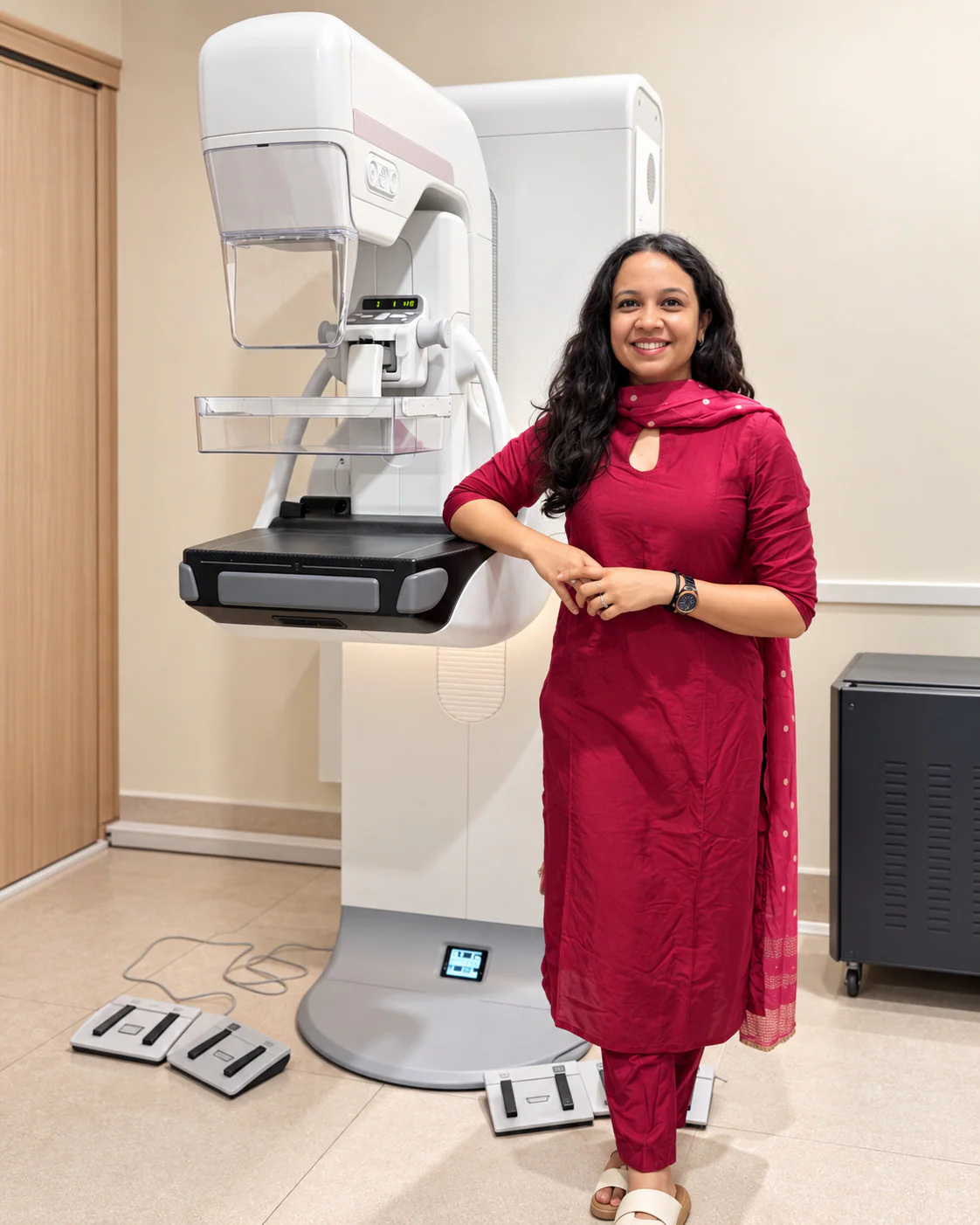
- Specialist breast radiologist
- Mammography & tomosynthesis
- Breast imaging in Lucknow
Understanding the examination
A closer look at breast tissue
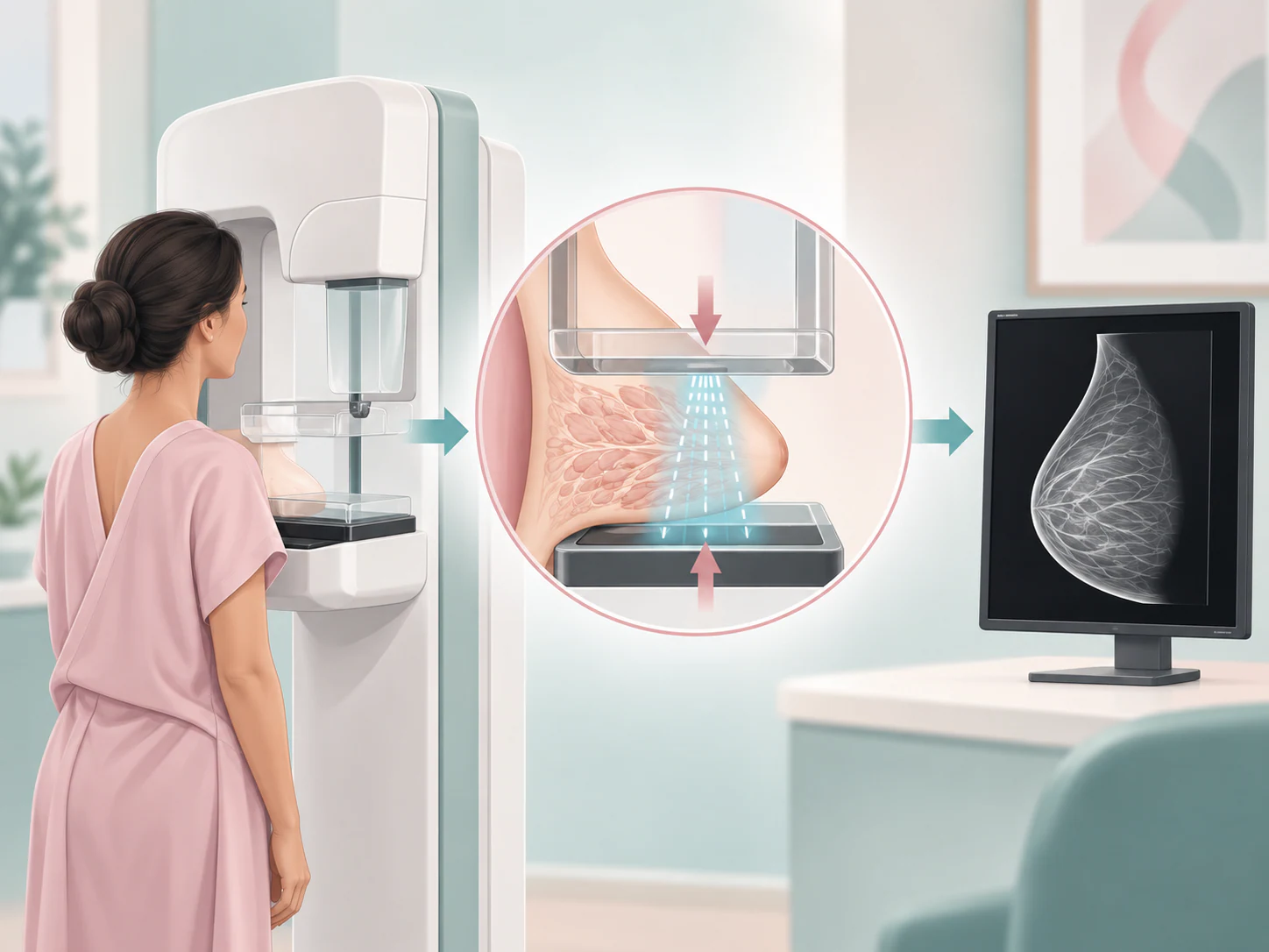
Mammography is an X-ray examination of the breast. It can show features of breast tissue that may need closer assessment. The radiologist reviews the images alongside your symptoms, history and earlier studies.
Screening and diagnosis
One examination, different reasons
The terms screening and diagnostic describe the purpose of mammography. Knowing which applies to your visit helps the team plan the examination and understand the question it needs to answer.
Screening mammography
Screening looks for breast changes in someone without a new breast symptom. Whether screening is appropriate, and when to repeat it, depends on a discussion of individual circumstances, history and the screening guidance being followed. There is no single schedule suitable for everyone.
Diagnostic mammography
Diagnostic imaging investigates a concern, such as a lump or nipple change, or takes a closer look at a finding from a previous examination. It may include additional views focused on a particular area. Tell the team about a new symptom even if your appointment was originally arranged for screening.

Reading the images
What can mammography help assess?
The radiologist evaluates the appearance, distribution and relationship of findings, rather than judging one feature in isolation. Comparing both breasts and reviewing earlier studies can help distinguish a longstanding appearance from a new change.
- Masses or areas of density
- An area may appear denser than surrounding tissue. Its shape and margins help guide whether closer assessment is needed.
- Calcifications
- Small calcium deposits can appear on a mammogram. Their pattern and shape matter; the presence of calcium alone does not establish a diagnosis.
- Architectural distortion
- This describes a change in the usual arrangement of breast tissue, sometimes without a clearly defined lump on the image.
- Asymmetry
- One area may look different from the corresponding region of the other breast. Further views may help explain overlapping tissue.
- Interval change
- A difference from an earlier examination can be important. Bring the actual previous images when possible, as a written report cannot show every visual detail.
Your experience
What happens during a mammogram?
Trained staff guide you through the examination and explain each position. Let them know about tenderness, restricted shoulder movement, recent surgery or anything that may make positioning difficult. You can ask questions throughout the visit.
Positioning
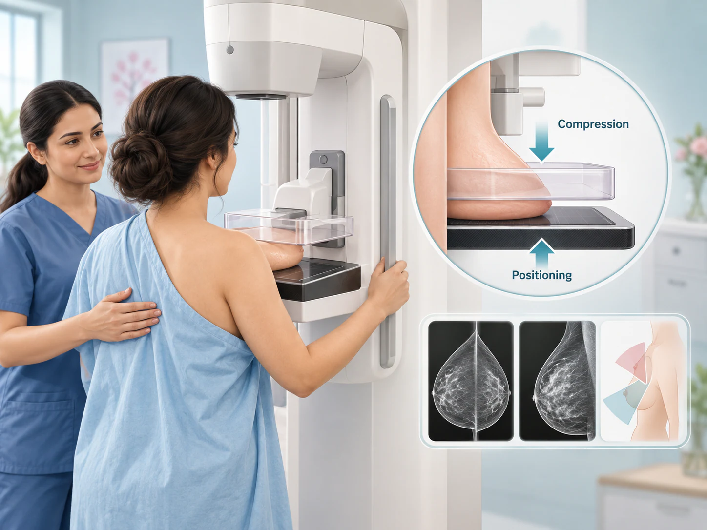
The breast is placed on the detector and carefully positioned to include the tissue needed for the image. You may be asked to adjust your posture or arm position.
Temporary compression
A paddle holds and spreads the tissue while the image is taken. Compression reduces movement and overlapping tissue, helping image clarity. It can feel uncomfortable; tell the staff if it is painful.
Views and image checks
Images are taken from the views appropriate to the examination. Compression is released between exposures. The team may need another position or a closer view before the imaging is complete.
A practical preparation checklist
What to bring and how to prepare
A little preparation can make it easier to answer questions and compare your images. Follow the instructions provided for your own appointment, especially if another examination is planned at the same visit.
For mammography in Gomti Nagar, contact Advanced Breast Care at Lucknow Imaging Centre. Tell the team whether you are arranging screening or have a symptom or referral; this helps plan the right type of visit.
Previous breast images and reports
Bring available mammograms, ultrasound or MRI records, including digital images or films. Mention where earlier examinations were performed if you cannot obtain them.
Referral and relevant history
Bring the referral when available. Tell the team about symptoms, previous breast procedures, treatment and relevant family history.
Pregnancy, lactation and implants
Tell the team before imaging if pregnancy is possible, if you are breastfeeding or if you have breast implants. These details help the team plan appropriately.
Practical clothing and instructions
Separate upper and lower garments can make changing easier. Check the appointment instructions about products used on the breast or underarm area. Ask about any preparation you are unsure of.
Do not stop prescribed medicines or change your routine on the basis of this page. If your appointment includes another procedure, ask for the instructions specific to that procedure.

Choosing the imaging technique
2D mammography and tomosynthesis
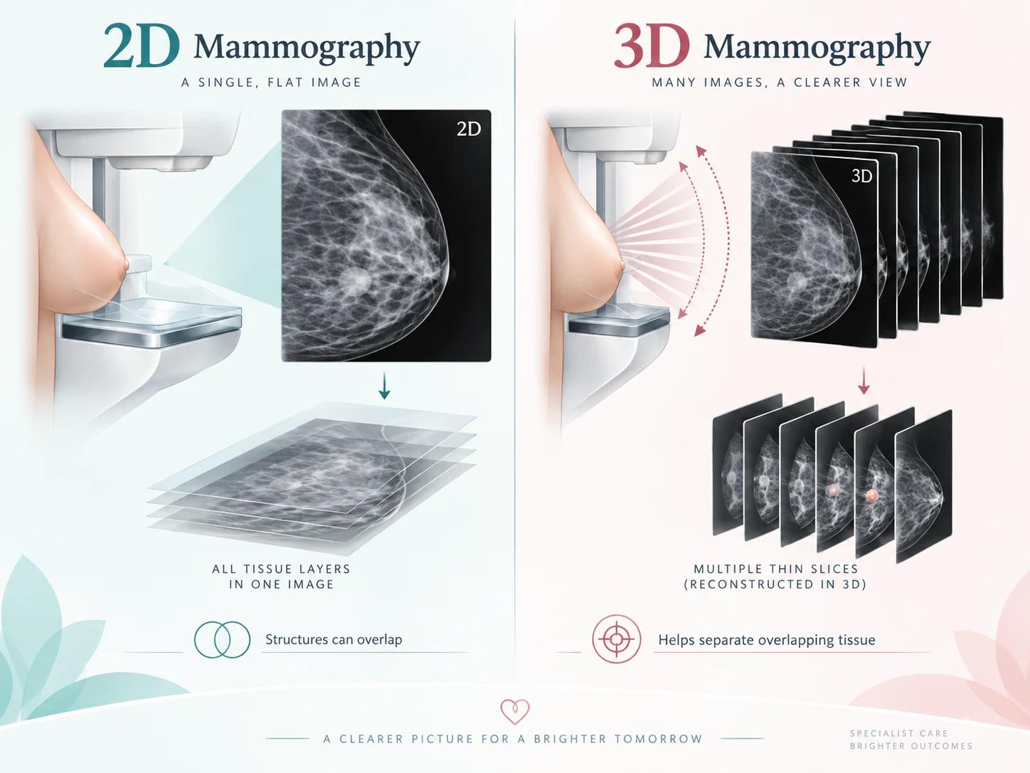
Conventional digital mammography creates two-dimensional images of the breast. Because tissue is projected onto a flat image, structures at different depths can overlap. The radiologist interprets those patterns using the available views and clinical information.
Digital breast tomosynthesis, also called 3D mammography, acquires projection images from different angles. A computer reconstructs these into thin sections that the radiologist can review through the breast. This provides another way to examine tissue that overlaps on a two-dimensional image.
The current equipment capability includes mammography and tomosynthesis. That does not mean every person needs the same examination. The technique is selected according to the clinical question and assessment; 3D imaging is not a guarantee that every abnormality will be detected.
Complementary information
Where breast ultrasound fits
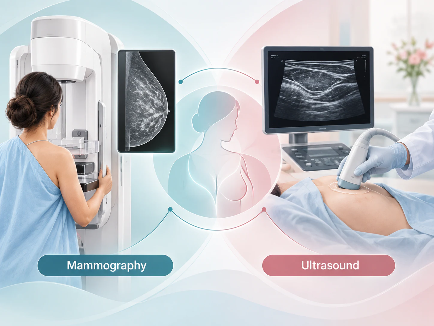
Ultrasound uses sound waves rather than X-rays. It can provide additional information about a particular area, including whether a finding contains fluid or appears solid. Mammography and ultrasound show tissue differently and answer different questions.
Ultrasound may be advised after a mammogram to examine a symptom or clarify an imaging finding. It is not automatically necessary for everyone, and it does not simply replace mammography. Your team considers the examination findings, symptoms and history when choosing additional imaging.
Reporting and results
From images to a clear next step
The radiologist reviews the images and, where available, compares them with previous examinations. The report records the findings, an assessment and any recommended next step. A recommendation can also depend on a symptom that needs attention even when the mammogram does not show an obvious explanation.
Ask how you will receive your report and who will discuss it with you. Keep a copy alongside the images for future comparison. If the report recommends another test or follow-up, clarify what is needed and how to arrange it with your care team.
A normal mammogram cannot exclude every breast cancer. New or persistent breast changes still deserve clinical assessment. Do not assume that an earlier reassuring result explains a new symptom.
If further evaluation is advised
More information does not automatically mean cancer
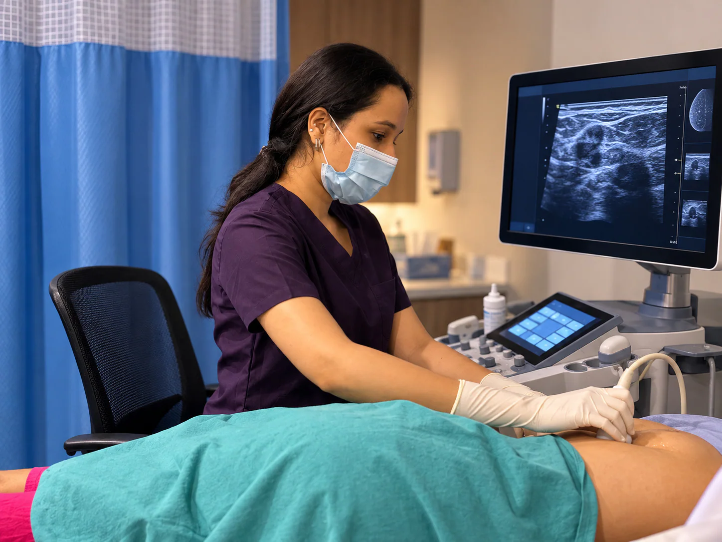
Additional mammographic views can help separate overlapping tissue or examine a small area more closely. Ultrasound may answer a different question about the same finding. Sometimes comparison with older images provides the information needed.
For selected findings, an interval imaging follow-up may be recommended to assess stability. When imaging cannot sufficiently explain a concerning area, a biopsy may be advised to obtain tissue for laboratory examination. These are different steps with different purposes, not an automatic progression for every patient.
Ask what the recommended test is intended to clarify and when the team wants it completed. An imaging assessment alone is not the same as a tissue diagnosis.
Putting terminology in context
Two terms you may see in your report
Breast density
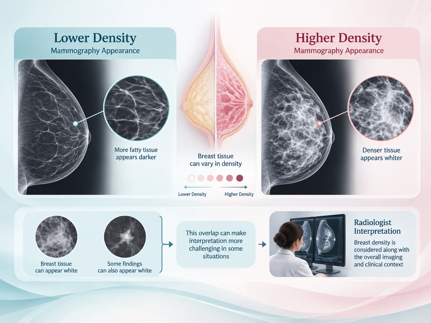
Density describes the relative appearance of fibrous and glandular tissue compared with fat on a mammogram. Dense tissue can make some findings harder to see because tissue and some abnormalities appear similarly white. Density is considered alongside other clinical information; it is not a personal risk score or a diagnosis by itself.
BI-RADS assessment

BI-RADS is a standardised system for describing breast-imaging findings and assessment. It helps communicate the result and the recommended next step. A BI-RADS category is not a cancer stage. Discuss the wording and recommendation with your care team rather than interpreting a category without its report.
Before and after your visit
Common questions about mammography
Is screening mammography different from diagnostic mammography?
Screening looks for changes without a new breast symptom. Diagnostic mammography investigates a symptom or an imaging finding and may need focused additional views. Tell the team why the examination was requested.
Is breast compression necessary?
Compression is part of mammography. It holds and spreads tissue during the exposure to help image quality. It is temporary and may be uncomfortable. Tell the staff about pain or positioning difficulties.
How long should I allow for the appointment?
The visit depends on the examination planned and whether additional views or assessment are needed. Ask the team how much time to allow when arranging your appointment.
Can I have a mammogram with breast implants?
Mammography can be performed in people with implants, but positioning and views may need adjustment. Tell the team about your implants when arranging the visit and before imaging begins.
Should I bring previous mammograms?
Yes, bring available previous images as well as their reports. Comparison helps the radiologist assess whether an appearance is longstanding or has changed.
Can mammography detect every breast cancer?
No. Mammography has limitations, including the effect of overlapping or dense tissue. A new or persistent breast symptom should still be discussed with your clinician, even after a reassuring mammogram.
Why might ultrasound be advised afterwards?
Ultrasound provides different information and may help assess a symptom or a specific finding. The need for it depends on the clinical question; everyone does not need both examinations.
What is 3D mammography?
Also called digital breast tomosynthesis, it uses projection images taken at different angles to reconstruct thin sections of the breast. Its use is selected according to the clinical context.
Does an abnormal mammogram always mean cancer?
No. A finding may have a benign explanation or may require more information. Additional views, comparison, ultrasound or other assessment can help establish the appropriate next step.
When might a biopsy be recommended?
A biopsy may be advised when tissue examination is needed to clarify a finding. Your team should explain the reason and the procedure being considered. A recommendation for biopsy is not itself a cancer diagnosis.
Specialist breast imaging in Lucknow
Need a breast imaging assessment?
Dr Pranjali Joshi is a breast radiologist and Lead Breast Imaging & Interventions at Advanced Breast Care at Lucknow Imaging Centre. The practice provides mammography and tomosynthesis capability for breast assessment.
Discuss the reason for your imaging visit with the team, including any new symptoms and previous examinations. Confirm the appointment arrangements and the records to bring.
Contact the practice