Breast symptoms · individual assessment
Breast Skin and Nipple Changes
Tell your clinician about a new or persistent change in breast skin or nipple appearance. Explain what looks different and when you noticed it. An examination and your history help determine whether imaging or further assessment is needed; appearance alone cannot identify the cause.
Contact the practice
Your usual appearance matters
What has changed?
Describe the change in your own words: what looks different, where it is and when it began. Mention whether it is new, persistent or becoming more noticeable.
Tell your clinician about any associated lump, discharge, pain or previous breast procedure. Appearance alone cannot identify the cause.
-
Where
Point out the part of the skin or nipple that has changed.
-
When
Note when you first noticed it and whether it is changing.
-
Other changes
Mention a lump, discharge, discomfort or other symptoms.
Describe the finding
A visible change is not a diagnosis
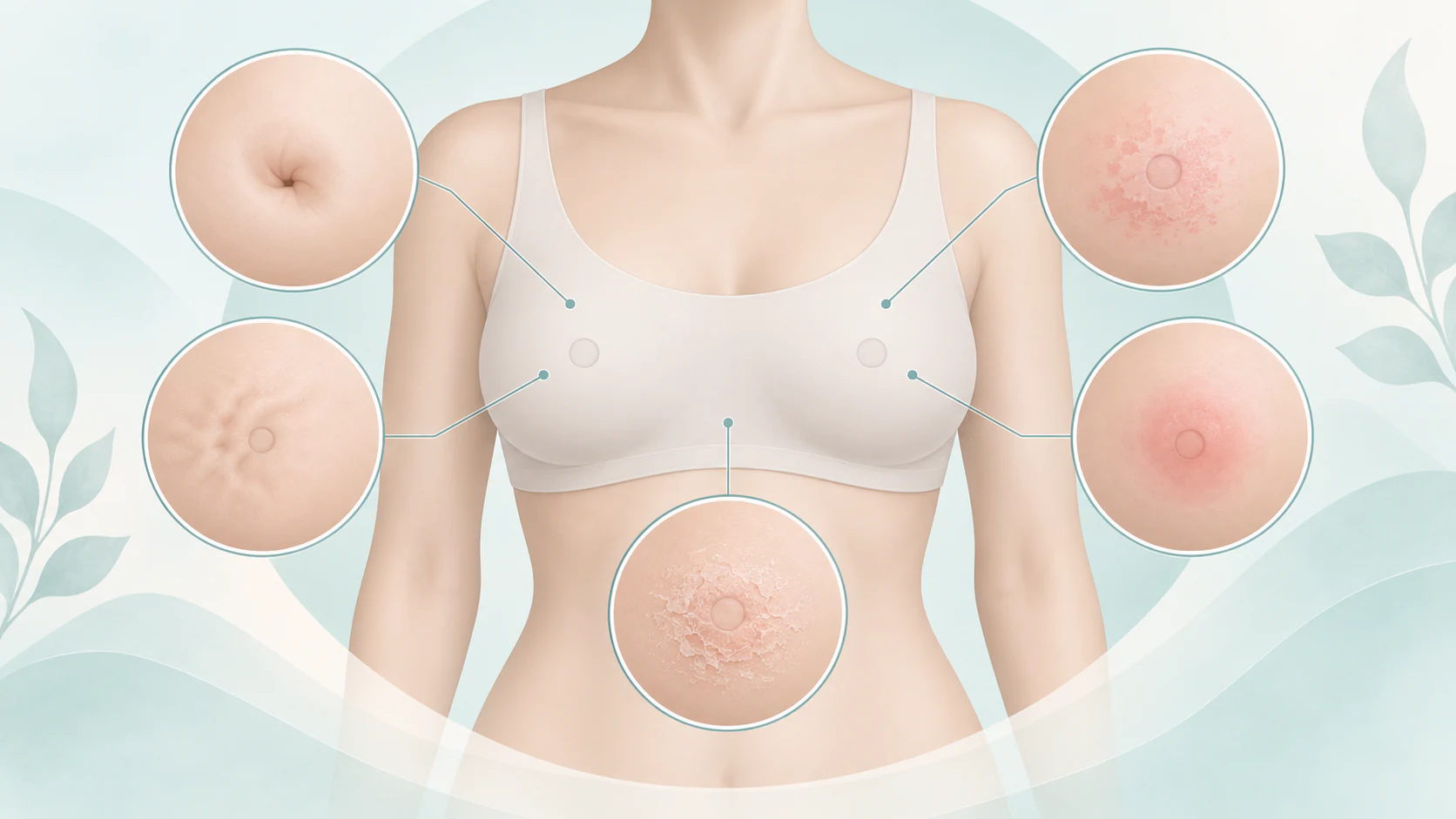
Nipple position, skin contour, texture and a persistent rash are all details that may be relevant to assessment. The examples below help describe a concern; they are not a visual cancer checklist and do not rank changes by seriousness.
-
Nipple position
Explain a new inward turn, pulling or change from the nipple’s usual position. Mention whether the appearance is longstanding or has recently changed.
-
Dimpling or a contour change
Describe a new indentation, pulling or difference in the skin surface. Point out the area without trying to identify a cause from its shape.
-
Persistent skin changes
Mention a rash, scaling, redness, swelling or a change in texture. Describe whether it persists, returns or is spreading, and whether there is discomfort.
Consider changes together
Mention what else you have noticed
Tell the clinician about any lump, discharge, pain or underarm concern alongside the visible change. Explain when each began and whether they appeared together. The absence of pain does not determine whether a visible change needs assessment.
-
A lump or thickening
Point out a new focal area as well as the visible change. One finding should not be assumed to explain another without assessment.
-
Discharge or discomfort
Describe discharge or pain and how it relates to the skin or nipple concern. Each detail can help the clinician understand the whole history.
History and examination
Clinical assessment gives the appearance context
A clinician may look at and examine the breasts and the area you have identified. Tell the team about earlier breast conditions, skin problems, biopsies, operations or other relevant procedures, including anything that changed the breast’s appearance.
Mention pregnancy or breastfeeding, medicines, relevant personal or family history and any previous assessment of the same concern. Explain what has changed since then. You can ask what the examination involves and discuss discomfort or practical needs before it begins.

Information that may help
Imaging can complement the examination
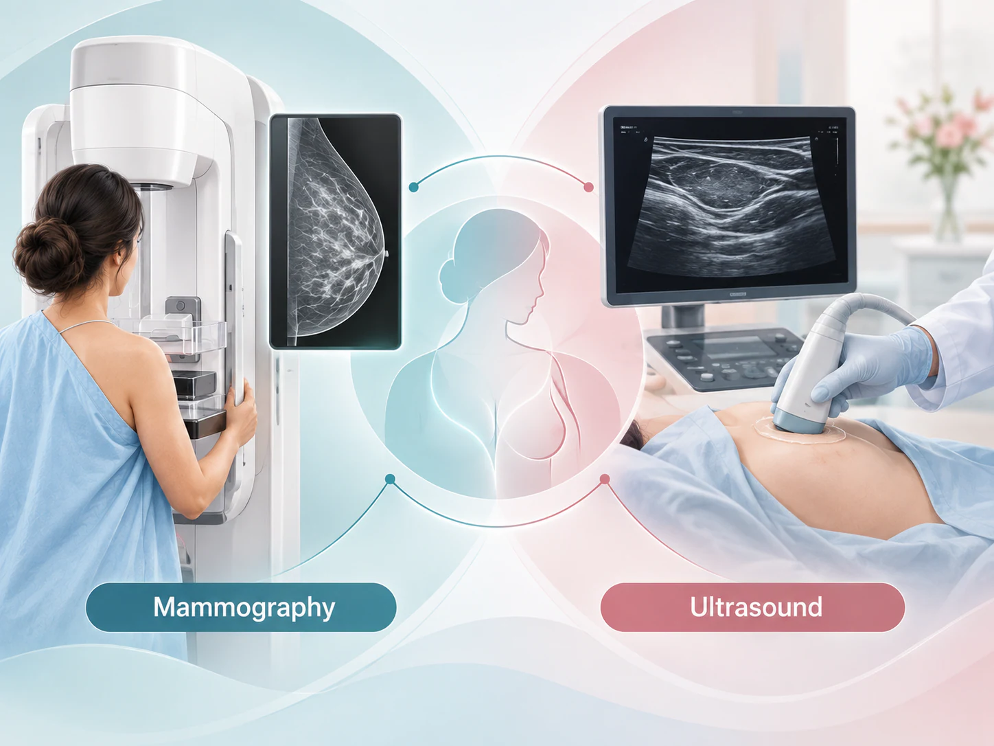
The clinical finding and your circumstances guide whether breast imaging is useful. Ultrasound may help assess an area of concern, while mammography can provide different information about the breast. One examination or a combination may be recommended when appropriate.
Imaging is one part of assessment; it does not replace looking at the skin or nipple and understanding the history. A persistent surface change may need further clinical evaluation even when an earlier imaging result was reassuring.
Assessment of a current symptom is different from routine screening. Tell the team about the visible change when arranging a visit so that the reason for the examination is clear.
When clarification is needed
Further assessment is selected for the finding
Some findings need additional imaging, comparison with previous studies or referral for further clinical assessment. Sampling may sometimes be recommended to clarify a skin, nipple or breast finding. It is not automatic for every visible change, and a recommendation does not itself mean cancer.
The method and the clinician involved depend on the finding being investigated. An image-guided breast biopsy and a sample of a surface skin change answer different questions; do not assume one procedure is suitable for every concern. Ask what is being assessed and who will explain the result.
Connect earlier care
Bring records that help explain the change
For assessment of breast skin or nipple changes in Lucknow, bring the information you already have. You do not need a complete set of records before raising a new concern. Tell the team about missing images or reports if comparison may be useful.
-
Earlier breast imaging
Bring mammograms, ultrasound images and written reports. Original digital image files may help comparison, so include them where available.
-
Procedure and medical history
Include relevant biopsy results, breast surgery or other procedure details, your referral and a medicine list. Explain whether a change followed previous treatment.
-
A note of the concern
Record what is different, when you noticed it and any associated symptoms. Bring the questions you want to discuss and confirm practical arrangements with the team.

A continuing conversation
Report a persistent or changing concern
Ask how the findings relate to the visible change, what remains uncertain and whether follow-up is needed. Clarify who will explain any outstanding results and what to do if the expected communication does not arrive.
If the change persists, returns or develops further after a reassuring assessment, update your treating clinician. A normal earlier scan does not explain every new skin or nipple concern. If you develop a red, hot or swollen breast with fever or feel acutely unwell, seek prompt medical assessment rather than waiting for a website enquiry.
Your questions
Common questions about breast skin and nipple changes
Does a nipple change always mean cancer?
No. Visible changes can have different explanations. A new or persistent change should be assessed because appearance alone cannot establish its cause.
Can nipple inversion be assessed with imaging?
Imaging may contribute, depending on the history and examination. Tell the clinician whether the position is longstanding, new or changing; that detail alone does not select the test.
What should I do about a new breast skin change?
Arrange a clinical assessment and describe what is different from your usual appearance. Mention when it began and any associated lump, discharge or discomfort.
Does breast redness always mean something serious?
Redness can have different causes and should be considered in context. A red, hot or swollen breast with fever or feeling unwell needs prompt medical assessment.
Can ultrasound assess skin or nipple-related concerns?
Ultrasound may help evaluate an area of concern when clinically appropriate. It complements the history and examination and cannot explain every visible surface change on its own.
Can mammography also be needed?
Yes, in some circumstances. Mammography and ultrasound can provide different information, and the clinical team decides whether one or both would help.
Does every visible change need a biopsy?
No. Sampling is considered when it is needed to clarify a finding. The appropriate method depends on what is being assessed; it is not the same for every skin, nipple or breast concern.
Should I bring previous breast images?
Yes, where available. Include the images and written reports, as well as relevant biopsy, operation or procedure records. These can help the team understand earlier findings.
What if earlier imaging was reassuring?
Tell your clinician about a new, persistent or changing concern despite that result. Earlier imaging is useful context, but it does not replace assessment of the current change.
Patient information sources
This guide supports a discussion with your treating team and does not replace an individual assessment.
Preparing for a discussion
Bring the story of the visible change
Explain what is new or different for you, describe any associated symptoms and bring relevant earlier records. The aim is an individual assessment and a clear explanation of the next step.
Contact the practice