Breast imaging in Lucknow · Dr Pranjali Joshi
Breast Clip and Marker Placement
A small breast clip or marker records the location of a finding or sampling site. It helps the team recognise that area on later imaging or when planning treatment. The reason for placing a marker should be explained before the procedure.
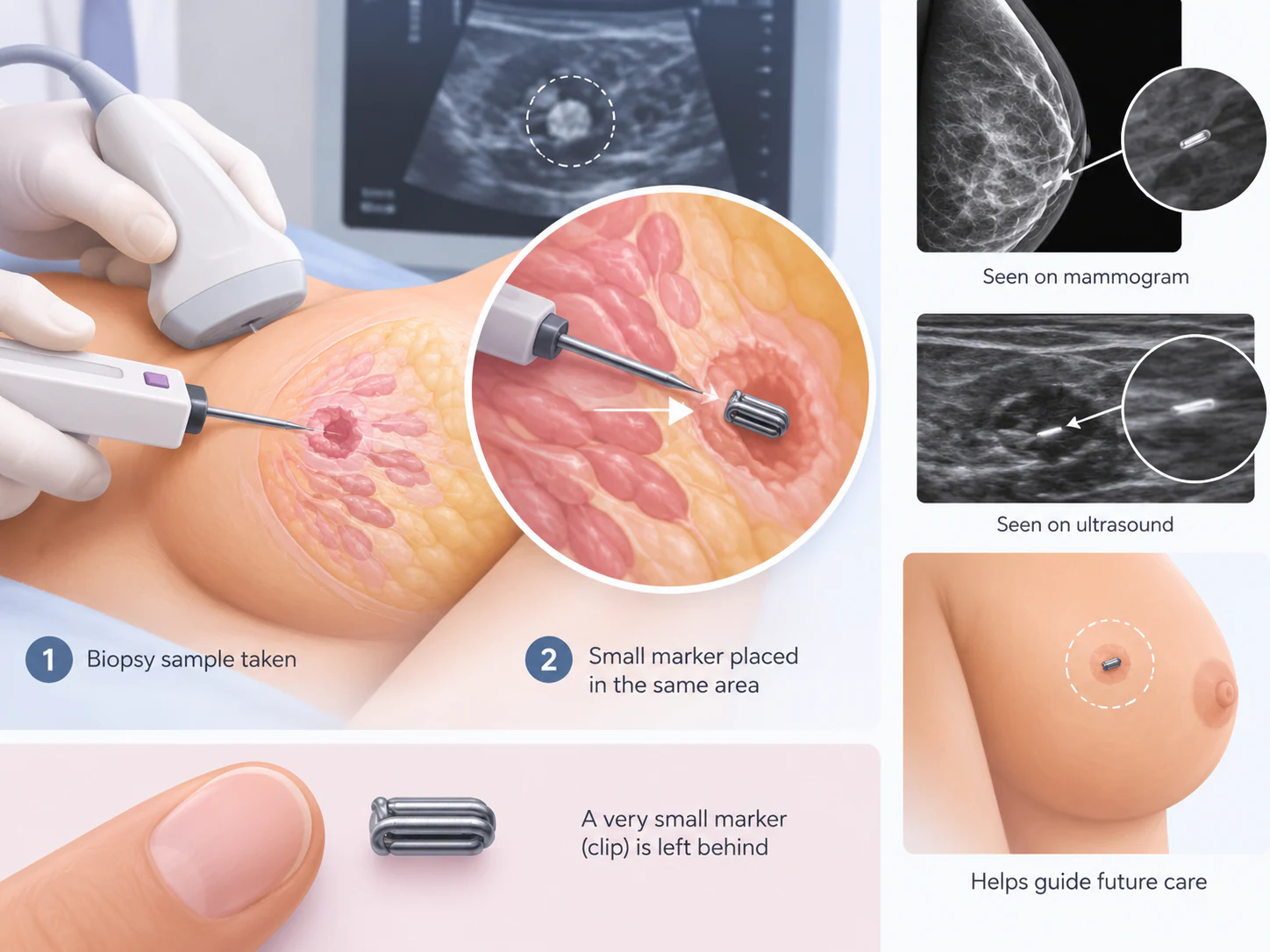
- A record of a particular site
- An individual clinical decision
- Information for future imaging
Start with its purpose
A marker records a location, not a diagnosis
A marker records where a finding or sampling site is located. It can help the team recognise the same area at a later visit.
The marker itself does not tell you the diagnosis. Imaging, sample results and your medical history still need to be reviewed together.
The site
Where the marker was placed and which finding it relates to.
The assessment
What imaging and any tissue results mean, and what the treating team recommends next.
An individual choice
Why placement may be discussed around a biopsy
A breast biopsy marker clip may be discussed when preserving a reference to the sampled area would help future care. It may also be considered before treatment that could change how a finding looks. These are examples of its purpose, not a recommendation for every patient.
The decision depends on what is being assessed, whether the site will need to be located again and the care plan. A previous biopsy without a marker does not automatically mean anything was missed. Conversely, placement of a marker does not mean that an operation has already been decided.
Ask why a marker is being proposed now and what information it will provide later. If you have more than one finding or have had previous procedures, the relationship between the new marker and earlier sites should be clear in the records.
Before placement
Bring the history of the site being marked
Preparation starts with understanding the proposed procedure. Clarify whether marker placement is planned during a biopsy or as a separate visit, which breast finding is involved and what imaging guidance is expected. These details affect the instructions you need.
Share your medicines, supplements, allergies and relevant medical history, including bleeding problems or previous reactions during procedures. Mention any possibility of pregnancy. The clinician can then decide whether further checks or tailored instructions are needed; do not stop prescribed treatment independently.
Imaging and procedure records
Bring earlier images and reports, biopsy results and details of any existing breast markers where available. Written reports alone may not show everything needed to identify a site.
Questions about the device
Ask how the marker will be documented, which product information you should keep and who can answer future questions about it.
Confirm the appointment location and any preparation directly with the team. If placement accompanies another procedure, ask which instructions apply to the combined visit rather than assuming that marker placement has a separate recovery plan.
The visit
Imaging guides placement and records the position
The team explains the reason for placement, checks the target and discusses consent. Imaging guidance is chosen according to the finding and clinical plan. The illustration shows an example; it does not establish which device or guidance method will be used for you.
Identify the intended site
The clinician reviews the relevant imaging and confirms the area to be marked. Let the team know about earlier procedures or markers.
Place the marker
The area is numbed as appropriate and the marker is introduced by the clinician using imaging guidance. Tell the team if you experience unexpected discomfort.
Document the relationship
Further imaging may be used to record the marker position relative to the target. Ask how this record will be shared with the clinicians involved in your care.
After the procedure
Get advice for your actual visit
Tenderness, bruising or bleeding can occur after placement, particularly when it is part of a biopsy. Infection and reactions to materials or local anaesthetic are also considerations to discuss. Your team should explain relevant risks without promising that every patient will have the same experience.
Follow the instructions you receive about the dressing, activities and any medicines. Those instructions should reflect all procedures performed at the visit. A general explanation of marker placement cannot replace an individual aftercare plan.
Ask whom to contact if discomfort worsens or you notice concerning swelling, bleeding, redness or other unexpected symptoms. Do not assume a new symptom is caused by the marker without assessment. If you feel acutely unwell, seek urgent medical care.
Keep the record
Help future teams identify the same site
Keep your procedure report with your breast imaging and pathology records. Useful information includes the date, which breast was marked, the site and the marker identification supplied by the team. If you receive a device card or product record, retain it rather than relying on memory.
Tell future imaging teams that a marker was placed, even if the procedure was performed elsewhere. Access to the earlier images and reports helps them connect the reference point with the original finding. A marker does not replace the history of why the area was sampled or assessed.
If you do not have the details, ask the centre that placed the marker for the relevant record. Avoid guessing the product from its appearance in a diagram or from another person’s device information.
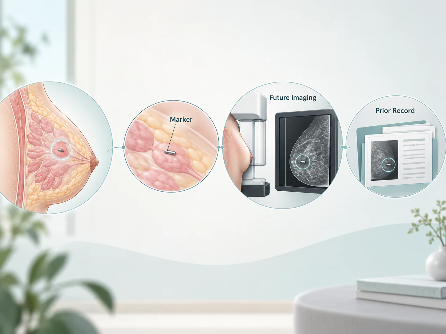
Device details matter
Questions about your marker and future scans
Markers can differ in design, material and the conditions attached to their use. This guide cannot confirm MRI suitability for an unidentified product. Before an MRI examination, tell the imaging team about the marker and provide the available identification and procedure records so they can perform the appropriate safety checks.
Ask the placing team whether the marker is expected to remain in place and whether a future procedure may remove it with the relevant tissue. Do not assume that all markers are permanent, all need removal or all behave in the same way on every imaging examination.
Questions about discomfort, device material or a possible change in position should be directed to the clinical team. Confirmation imaging and the written placement record provide context that a general website cannot supply.
Related but distinct
A marker clip is different from a localisation wire
A marker records a site for reference. Wire localisation helps identify a target for a planned operation. A person with a marker may still need localisation if surgery is later recommended; the team chooses how to locate the relevant area.
Neither device substitutes for the other automatically. Ask whether the recommendation is to document a site, guide an operation or both at different stages. This distinction can make referral letters and discussions with different teams easier to understand.
Next steps
Separate the placement record from the biopsy result
The placement record describes the marker and its relationship to the intended site. If a biopsy was also performed, its pathology report answers a different question about the tissue. Successful marker placement is not the same as receiving a diagnosis.
Confirm who will discuss any biopsy result and what follow-up is planned. If the marker was placed to support a treatment pathway, the responsible treatment team should explain the next stage. Do not infer a treatment decision from the presence of the marker alone.
For future appointments, bring these records together rather than presenting a marker card without the associated imaging history. The goal is a clear connection between the site, the assessment and the agreed care plan.
Your questions
Common questions about breast clip and marker placement
Does a breast marker clip mean I have cancer?
No. A marker identifies a site, not a diagnosis. The meaning of the finding depends on imaging, clinical assessment and any biopsy result.
Is a marker always needed after a breast biopsy?
No. The decision depends on the finding and whether recording the site is useful for future care. Ask why placement is or is not being recommended for you.
Is marker placement a treatment for the finding?
No. Placement records a location. It does not remove the finding or replace the assessment and any treatment recommended by your clinical team.
Can different patients have different markers?
Yes. Marker designs and materials can differ. Keep the product or procedure information supplied for your own marker rather than using another patient’s details.
Can I have an MRI after marker placement?
Tell the MRI team about the marker and provide its identification details. They must check the conditions for the actual device; this page cannot give blanket MRI clearance.
Will the marker stay in place forever?
That depends on the marker and care plan. Ask whether it is expected to remain or may be removed with tissue during a later procedure. Removal is not automatically required just because a marker is present.
What should I keep for future appointments?
Keep the placement report, prior images, relevant biopsy results and any device identification record. Tell the next imaging team which breast and site were marked.
Will I still need wire localisation if I have a clip?
Possibly, if surgery is planned and the area needs localisation. A clip and a wire serve different purposes, so the surgical and radiology teams decide what is appropriate.
What if I have discomfort after placement?
Follow your aftercare instructions and contact the treating team about worsening pain, swelling, bleeding or other concerns. Do not assume the marker explains a symptom without assessment.
Your next step
Know why the site is being marked
When discussing breast clip or marker placement in Lucknow, ask about its purpose, the imaging record and any device-specific information to keep. Dr Pranjali Joshi’s breast imaging and interventions practice considers the finding in its clinical context. The next step should follow your individual assessment and care plan.