Breast symptoms · individual assessment
Breast Lump Assessment in Lucknow
A new breast lump or an area of thickening needs assessment. How it feels cannot tell you its cause. Your doctor starts with your history and an examination, then considers imaging or sampling if more information is needed.
Contact the practice
Understanding a change
A lump can have different explanations
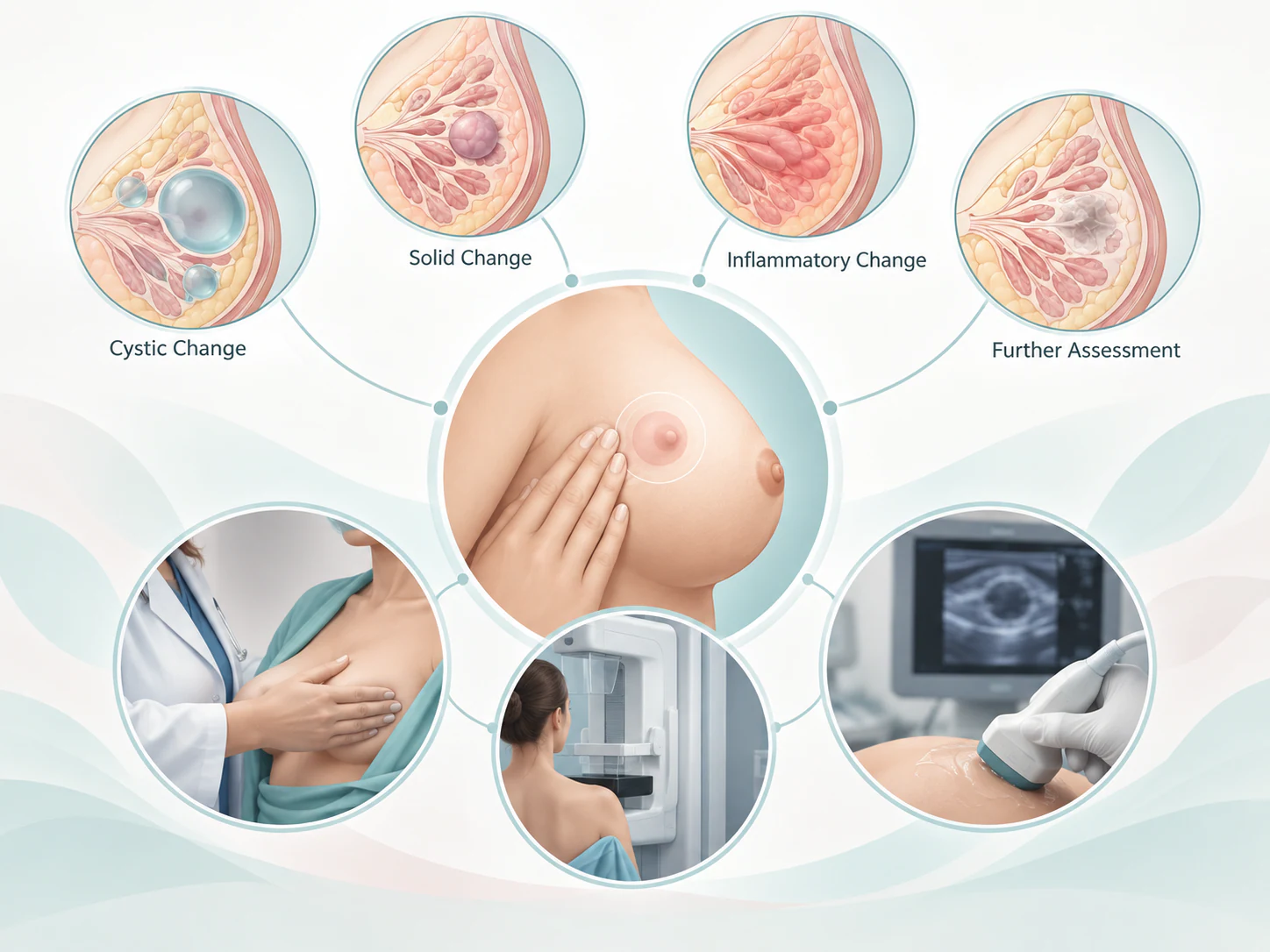
A breast lump may relate to a fluid-filled cyst, a non-cancerous tissue growth or another change in breast tissue. Some findings need further investigation to determine their cause. These possibilities cannot be reliably distinguished by comparing your breast with a picture or description.
A new lump, a persistent focal area or a change in a previously assessed lump deserves a clinical discussion. Pain, softness or movement alone should not be used to decide that a lump is harmless or serious. Explain what you have noticed without trying to give it a medical label.
Start with your experience
What to tell your doctor
Tell your clinician where the lump is, when you first noticed it and whether it has changed. Mention other symptoms and whether the same area has been assessed before.
Bring any earlier breast images, reports or procedure records. Your history and an examination help the clinician decide whether imaging or sampling could explain the finding.
Tests chosen for the question
Imaging adds information to the clinical assessment
The choice of imaging depends on age, the examination finding, pregnancy or breastfeeding, previous studies and the referral question. A recommendation is tailored to that context; this page is not a tool for choosing your own test.
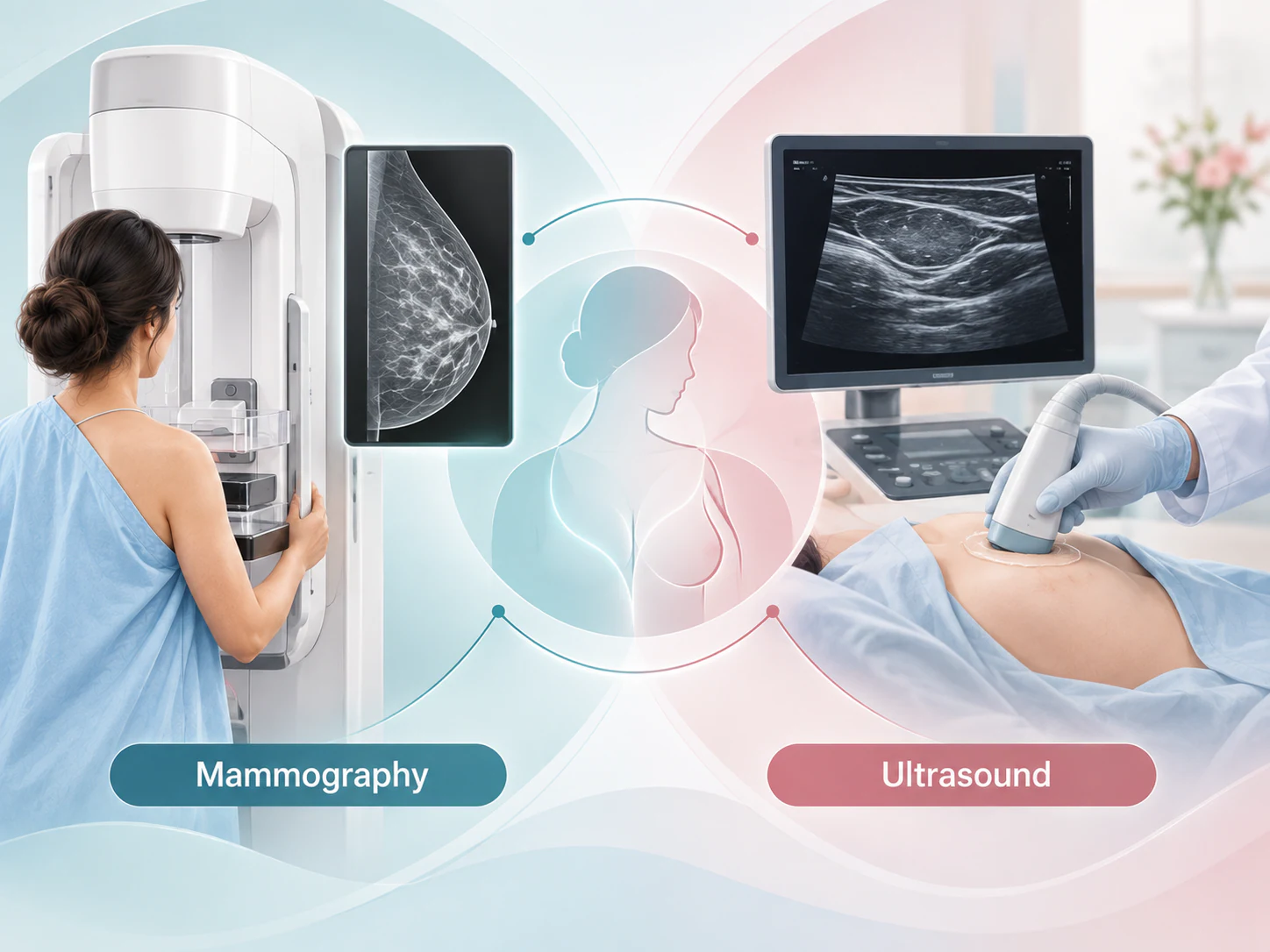
Targeted ultrasound can examine the area of concern and help describe whether a finding contains fluid, solid tissue or both. It cannot settle every diagnosis. Mammography or tomosynthesis may contribute different information about the breast and the finding under review.
Mammography and ultrasound can complement one another. Sometimes both are recommended; neither is universally better for every lump. Let the team know exactly where you feel the change so the imaging can be considered alongside the clinical finding.
For younger women, ultrasound is often the first imaging examination for a breast lump. Age alone cannot establish the cause, and a persistent concern still needs review even when an earlier scan was reassuring.
When more information is needed
Sampling is considered when it can clarify a finding
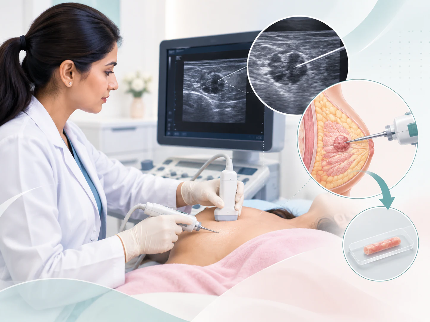
Not every breast lump needs a biopsy, FNAC or surgery. Sampling may be recommended when the clinical and imaging information needs laboratory clarification. A recommendation to take a sample is not itself a diagnosis of cancer.
Core biopsy obtains small tissue samples. Fine needle aspiration cytology (FNAC) obtains cells or fluid for cytological examination. The clinical question and the type of finding help determine which method is appropriate; the procedures are not interchangeable in every situation.
Ask why a sample is being recommended, how to prepare and who will explain the result. Laboratory findings are assessed with the examination and images. If the information does not fit together, further review or sampling may be discussed.
Bring earlier information
Earlier records can help connect the findings
For a breast lump assessment in Lucknow, bring relevant records that you already have. Images and written reports serve different purposes, so include both where possible. Tell the team if any records are missing rather than postponing a discussion while trying to assemble a complete file.
-
Breast images and reports
Bring previous mammograms, ultrasound images and their reports, including original digital image files where available. Include the dates and the place where earlier examinations were performed.
-
Sampling and procedure records
Include earlier biopsy or cytology reports, operation records and any breast marker information. Mention which side and area were previously assessed.
-
The story of the change
Note when the lump appeared, where it is and what has changed. Bring the referral and a list of your questions, medicines and relevant medical information.
-
Practical arrangements
Confirm the visit location and any examination-specific preparation with the team. If travelling, check the arrangements before booking transport; this page does not establish a same-day sequence of tests.

Results in context
The next step depends on what the assessment shows
Possible outcomes include reassurance with advice, comparison with previous images, additional imaging, planned follow-up or sampling. Some findings need a discussion with the treating team about further care. These are possible directions, not a fixed pathway that everyone must complete.
Ask how the result explains the lump you feel, whether anything remains uncertain and what follow-up is recommended. Clarify who will communicate the results and how to follow up if that communication does not arrive.
If a lump persists or changes despite reassuring earlier imaging, tell your treating clinician. An earlier normal result does not replace assessment of the current concern. When clinical, imaging and laboratory findings do not agree, the team may need to review the information together.
Your questions
Common questions about breast lump assessment
Does a breast lump always mean cancer?
No. A lump can have several causes, including non-cancerous changes. A new or changing lump needs assessment because touch alone cannot establish its cause.
Which test is used to assess a breast lump?
The clinician considers your history, examination, age and other relevant circumstances before recommending imaging. There is no single examination that answers every breast lump question.
Can ultrasound tell what a lump is?
Ultrasound can help describe the finding, including whether it is fluid-filled, solid or mixed. Some findings still need comparison, follow-up or sampling to clarify their meaning.
Can mammography and ultrasound both be needed?
Yes. They can provide complementary information. The recommendation depends on the finding and clinical context, rather than one test being universally better.
Does every breast lump need a biopsy?
No. Sampling is advised when it is needed to clarify a finding. The team considers the clinical and imaging information and explains why a particular method may be useful.
What records should I bring?
Bring earlier breast images and reports, any biopsy or cytology results, relevant procedure and marker details, and your referral. Original image files can be useful for comparison.
What if earlier imaging was normal but I still feel a lump?
Tell your treating clinician that the concern persists or has changed. The current finding needs to be considered alongside the earlier result, and further assessment may be needed.
Can a lump be assessed during pregnancy or breastfeeding?
Yes. Tell the clinician that you are pregnant or breastfeeding so that assessment and any imaging can be planned appropriately. Do not assume that these circumstances explain a new lump without assessment.
What if examination, imaging and sampling results do not agree?
The team reviews the findings together rather than relying on one result in isolation. Further discussion, comparison, imaging or sampling may be recommended to resolve the uncertainty.
Patient information sources
This guide supports a discussion with your treating team and does not replace an individual assessment.
Preparing for a discussion
Bring the story of the change to your assessment
Describe the change in your own words, bring the records you have and note the questions you want answered. The aim is an individual assessment and an understandable next step.
Contact the practice