Breast imaging in Lucknow · Dr Pranjali Joshi
3D Mammography in Lucknow
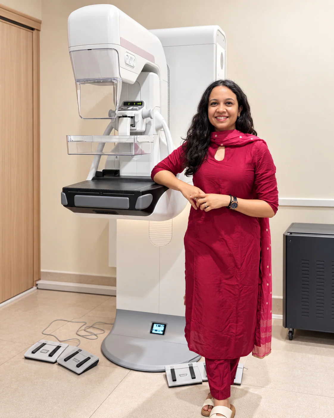
3D mammography takes breast X-ray images from different angles and combines them into thin sections for the radiologist to review. It is also called digital breast tomosynthesis. Whether it is useful for you depends on the reason for your examination.
- Reconstructed section review
- Screening or diagnostic context
- Compared with your clinical history
Understanding the examination
Looking through layers of breast tissue
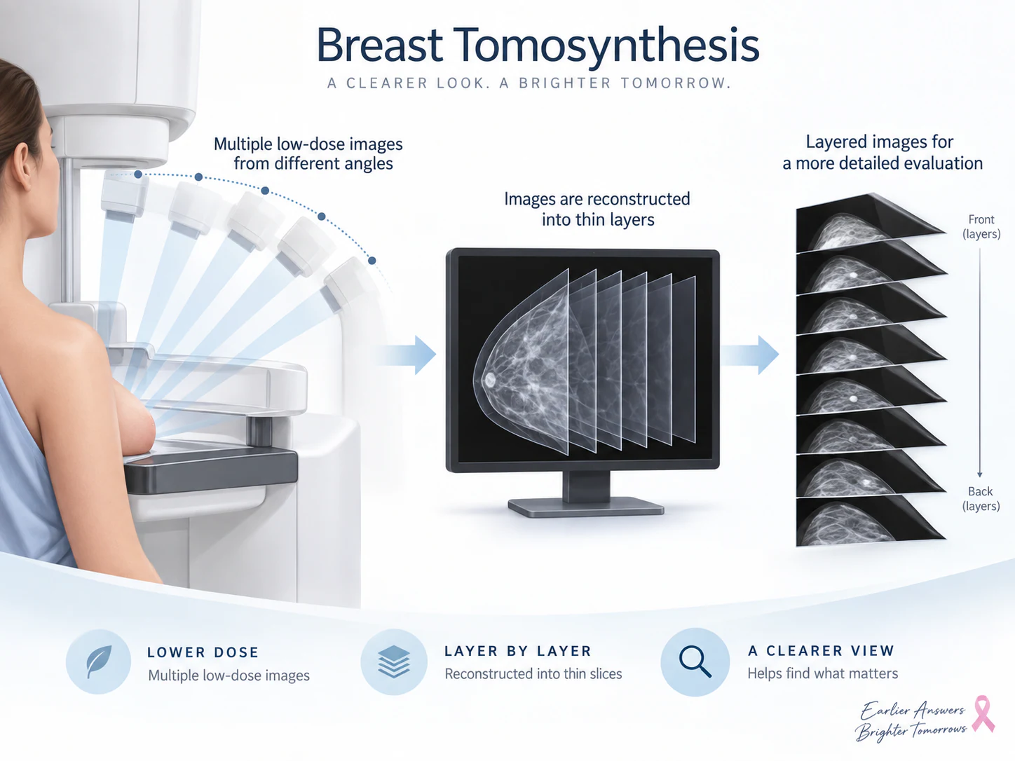
During tomosynthesis, the equipment takes breast X-ray images from different angles. A computer combines them into thin sections that the radiologist can review one by one.
This can help separate overlapping tissue. The images still need to be interpreted with your history and any other relevant tests.
Two ways of reviewing tissue
How do 2D and 3D mammography differ?
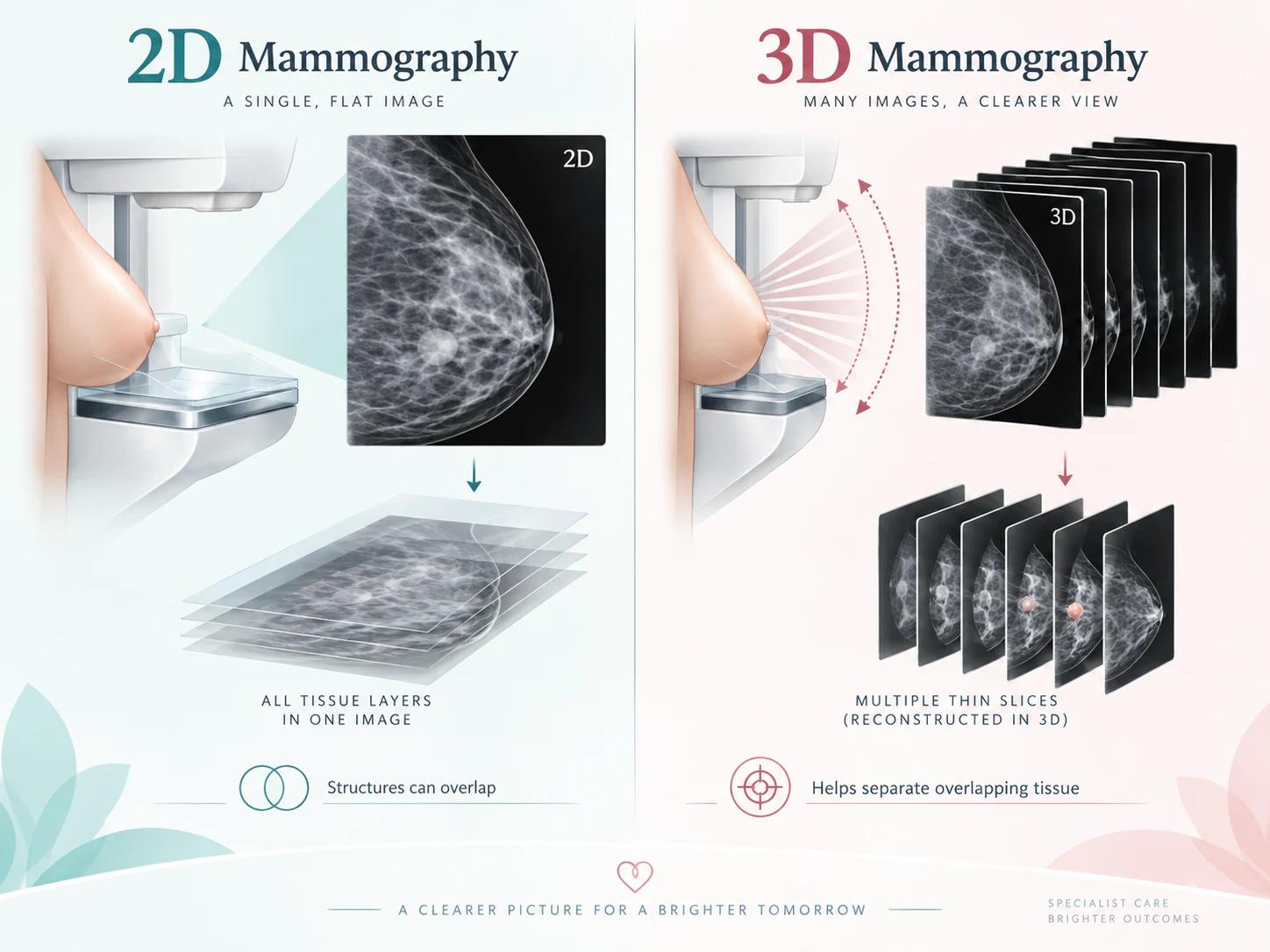
The distinction is principally how the image information is acquired and displayed. Conventional 2D mammography gives a projection view; tomosynthesis adds a reconstructed section series. The word “3D” does not mean a scan is free of limitations or that another examination will never be needed.
A 2D projection
Structures at different depths contribute to the same view. The radiologist examines the overall pattern and any area that needs closer attention.
A reconstructed section series
The radiologist can move through sections to examine how an area appears at different depths, reducing some of the difficulty caused by superimposed tissue.
The appropriate approach depends on the imaging question and available prior information. It is not a choice patients need to make from a technology label alone.
Choosing an examination
When might tomosynthesis be considered?
Tomosynthesis may be used in a screening setting or as part of a diagnostic assessment when appropriate. A radiologist may consider it to examine tissue overlap or look more closely at an area that is difficult to interpret on a projection image. The decision is made alongside the history and other findings.
Tell the team if the appointment relates to a new symptom, even if the booking was originally described as screening. Screening and investigation of a specific concern ask different questions. A new lump or persistent focal concern needs to be brought into the assessment rather than left out because an imaging appointment is already arranged.
An examination plan can change as information becomes available. Additional views or a different imaging method may be useful; this does not mean every patient will have the same combination of tests.
Breast density
Density provides context, not a diagnosis
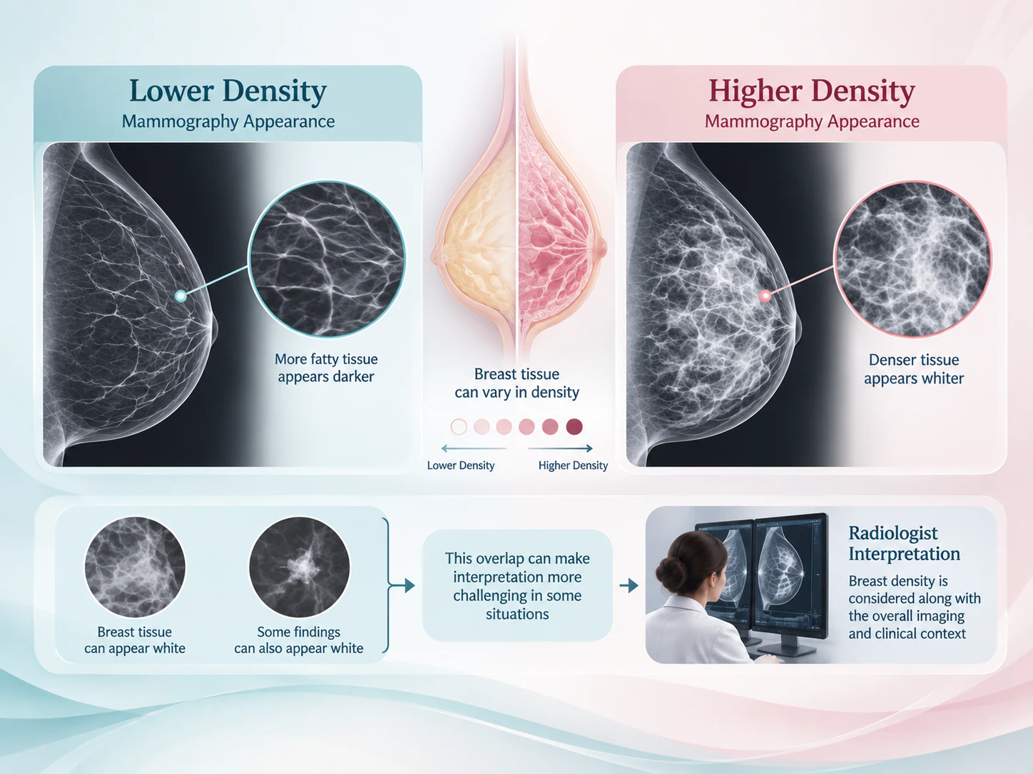
Breast density describes the relative appearance of fibrous and glandular tissue and fat on a mammogram. Dense tissue and some abnormalities can both appear light on X-ray images, which can make interpretation more difficult. It is an imaging description, not something reliably determined by how a breast feels.
Tomosynthesis can help separate overlapping appearances, but it does not remove every challenge associated with dense tissue. A density statement alone should not be treated as a diagnosis or an automatic instruction to have every available test. Ask how it affects interpretation of your particular examination and whether anything further is recommended.
Your visit
What happens during the examination?
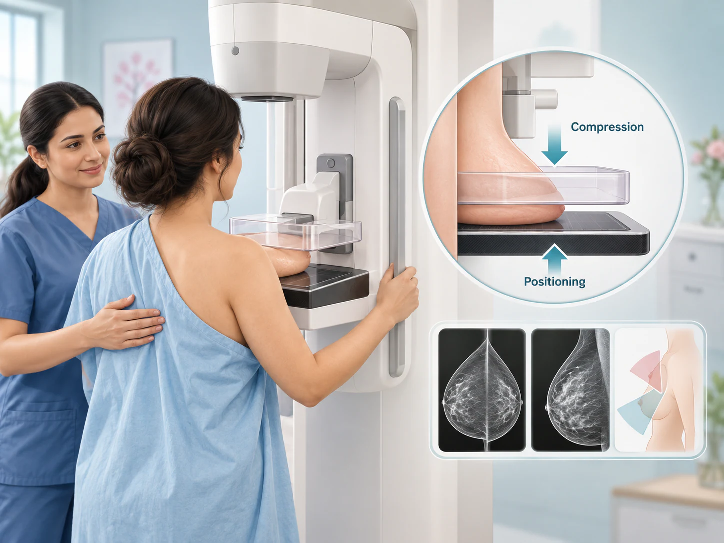
The experience is similar to mammography in several practical respects. The breast is positioned on the equipment and held with temporary compression while images are acquired. For tomosynthesis, the X-ray tube moves to collect projections from different angles; reconstruction takes place digitally afterwards.
Compression is still required. Tell the team about tenderness, difficulty standing or positioning, previous surgery or implants before the images are taken. If you experience discomfort, communicate with the person carrying out the examination so they can respond appropriately.
Before the images
The team reviews the reason for the visit and positions you for the requested views. You can explain any concerns about comfort or movement.
During acquisition
Follow the team’s instructions while the breast is held briefly in position. The equipment collects the projections needed for reconstruction.
After acquisition
The images are checked for the information needed. Sometimes an extra view is requested before the examination is complete.
Preparing for assessment
Bring the history as well as the images
Bring earlier mammograms, ultrasound images and reports where available, together with the referral and details of previous breast procedures. Actual images can help comparison in a way that a written summary alone cannot. If records are held elsewhere, ask how you can obtain them before the visit.
Mention any possibility of pregnancy before an X-ray examination. Follow the preparation instructions supplied for your appointment, including advice about products on the breast or underarm area. Tell the team about the symptom that prompted the referral and where you have noticed it.
The practice in Lucknow can confirm the requested examination and arrangements using the contact details supplied with your referral. Ask about availability before travelling; this page does not promise appointment slots, fees or a particular report turnaround.
If you have been advised to have digital breast tomosynthesis or a 3D mammogram in Lucknow, share the referral when arranging your visit. Confirm the examination and preparation before attending the Gomti Nagar practice.
Complementary information
Why ultrasound or another assessment may still be useful
Tomosynthesis is a form of mammographic imaging. Ultrasound uses sound waves and provides a different view of tissue. When a particular area needs characterisation, the methods may contribute different information. Having tomosynthesis does not automatically remove the need for ultrasound, nor does ultrasound universally replace mammography.
The next step should answer the remaining question. It may involve reviewing prior studies, targeted ultrasound, further mammographic views or a planned follow-up. Sampling is considered only when indicated. Ask which finding the recommendation relates to and what information the proposed step is expected to provide.
Interpretation and next steps
Understanding your report
The radiologist reviews the reconstructed images together with other available views and earlier examinations. The report describes the assessment and any recommendation. Clinical symptoms and history remain important, including concerns that persist even when imaging has not identified an explanation.
Tomosynthesis uses X-rays and has limitations. It cannot identify every breast cancer, and an appearance that requires investigation is not necessarily cancer. Decisions about radiation exposure and further testing should be considered in relation to the individual clinical question rather than a promise that any test is completely risk-free.
Before leaving, ask how the report will be made available and who will explain the next step. If additional assessment is advised, keep the written recommendation and share it with your treating doctor. Do not assume that silence means a normal result; make sure you receive and understand the report.
Your questions
Common questions about 3D mammography
Is 3D mammography the same as tomosynthesis?
Yes. Digital breast tomosynthesis is commonly called 3D mammography. It collects projections from different angles and reconstructs a series of sections for the radiologist to review. The names refer to the same imaging approach.
How is it different from regular mammography?
A conventional 2D mammogram presents projection images. Tomosynthesis adds reconstructed section review, which can help assess tissue overlap. The choice of examination depends on the clinical question; neither description guarantees that no other test will be needed.
Is compression still required?
Yes. Temporary breast compression is part of the acquisition process. Tell the team about tenderness or positioning difficulties and speak up if you are uncomfortable during the examination.
Does everyone need 3D mammography?
No. The imaging plan depends on the reason for assessment, relevant history and radiologist’s judgement. Discuss what information tomosynthesis is expected to add in your situation rather than assuming it is a requirement for every visit.
Can it replace breast ultrasound?
Not universally. Ultrasound and mammographic imaging show tissue differently. Ultrasound may help answer a question that remains after mammography or tomosynthesis, and the need for it is considered individually.
Should I bring previous images?
Yes, if available. Bring actual images as well as written reports and details of earlier procedures. Comparison can help the radiologist understand whether an appearance is new or was present previously.
What if further imaging is recommended?
Ask which area needs clarification and what the proposed examination will add. A request for further imaging is not itself a diagnosis. Keep the recommendation and arrange the next step with your treating team.
Is it used for screening and diagnostic assessment?
It may be used in either setting when appropriate. Screening concerns assessment without a specific symptom; diagnostic imaging addresses a symptom or finding. Tell the team about any new concern so the examination reflects the reason for your visit.
Your next step
Prepare for an informed breast imaging visit
Gather your previous images, referral and questions. Dr Pranjali Joshi’s breast imaging practice in Lucknow considers the examination in the context of the concern being assessed. The next useful step is understanding why a test has been requested and how its result will be reviewed.