Understanding breast assessment
Breast Symptoms and Assessment
Noticed a lump, pain, discharge or another breast change? Start with the guide that matches your concern. Symptoms can have different causes. A clinician can review what you have noticed and advise whether imaging or further assessment is needed.
Explore the assessment pathway
Common reasons for assessment
Start with the change you have noticed
Choose the guide closest to the change you have noticed. These are reasons to seek advice, not a checklist for diagnosing yourself. Tell your clinician about a concern even if it does not fit one of these groups.
-

A lump or focal thickening
Describe where you feel the change and whether it is new or changing. A lump can have different causes; touch alone cannot determine what it represents.
-

Nipple discharge
Tell the clinician when you noticed discharge, whether it occurs without touching the nipple and whether it involves one or both breasts. Its cause cannot be settled from its appearance alone.
-
Nipple or skin changes
Mention a new change in nipple position, a rash, skin dimpling or a change in the breast’s appearance. Describe what is different from your usual appearance.
-
Focal or persistent pain
Explain where the discomfort is, how long it has been present and whether it changes. Pain has many possible explanations; an assessment can establish whether imaging would be useful.
-
Underarm swelling or another focal concern
Point out the area that worries you and any associated breast change. A clinician can consider the concern alongside your history and examination.
-
A finding on earlier imaging
A report may recommend further assessment even when you have not noticed a symptom. Bring the images and report so the reason for review is clear.
Describe it in your own words
What changed, where and when?
A useful starting point is the story of the concern: when you first noticed it, its location and whether it has persisted or changed. Explain any associated symptoms and whether the concern affects your daily activities. You do not need to decide whether it is a cyst, a tumour or another condition before attending.
Mention earlier breast imaging, biopsies, surgery or other relevant procedures. If a similar concern was assessed before, bring that information and explain what is different now. Notes made before the appointment can help you remember questions without having to repeatedly check the area yourself.
Tell the team about relevant medicines and medical conditions, pregnancy or breastfeeding, and personal or family history. The clinician can decide which details affect the assessment. This is a discussion about your individual situation, not an online risk score.
The reason for the visit
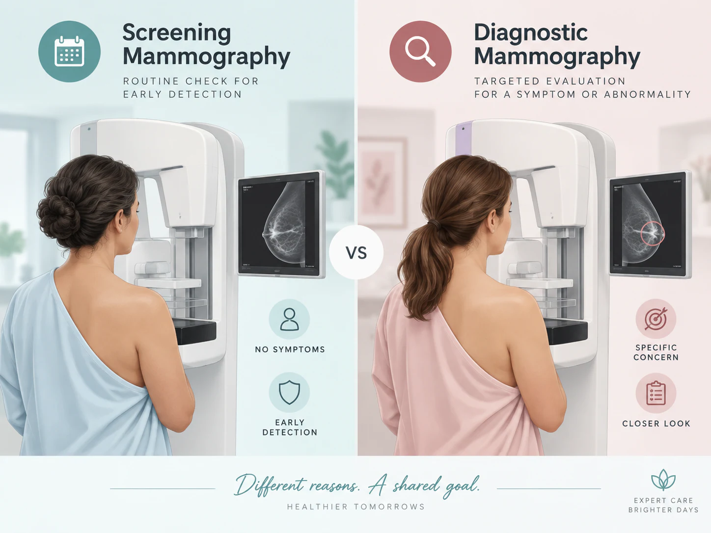
Symptom assessment is different from screening
Screening is planned when there is no new symptom. A symptom-led assessment addresses a particular change or concern. Let the team know about symptoms when arranging your visit; do not assume that a routine screening appointment is the right way to investigate them.
A previous reassuring screening result is useful information, but it does not explain every new change. If you notice something new or your concern continues, discuss it with your treating clinician. The appropriate response depends on the current situation.
The broader assessment pathway brings clinical information and images together. Not everyone needs the same examinations or every possible stage. Recommendations about routine screening and those about a current symptom should be explained separately.
An individual assessment
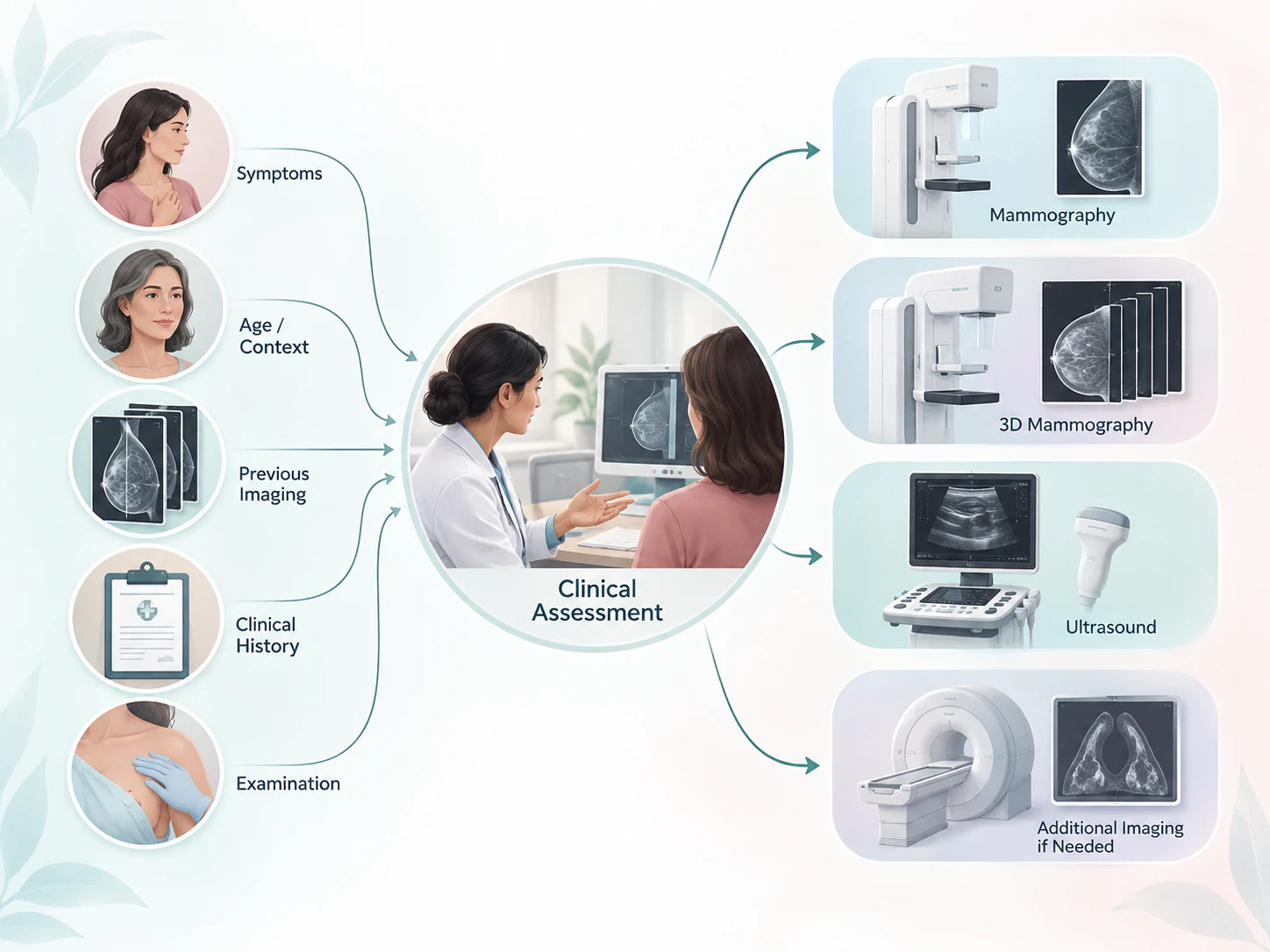
How your team chooses the next test
Your clinician considers the symptom, examination, earlier images and relevant history before recommending tests. Pregnancy, breastfeeding or a previous procedure can also affect the choice.
Dr Joshi contributes breast imaging and intervention expertise, with communication to your referring or treating clinician when needed. The illustration shows possible considerations, not a fixed sequence of tests.
How imaging may fit
Different examinations contribute different information
The team may recommend one examination or a combination. No modality is universally better for every breast concern. Use the service information to understand a recommendation and prepare questions, rather than to select your own investigation.
-
Mammography and tomosynthesis
Mammography uses X-ray images of the breast. Tomosynthesis provides reconstructed views that may contribute to an assessment. Their role depends on the clinical question and the finding being reviewed.
-
Breast ultrasound
Ultrasound can assess a particular area and help characterise a finding. It may complement mammography, so both may sometimes be needed. It is not an automatic replacement for another examination.
Completing the picture
Further assessment does not itself mean cancer
Additional views, a targeted ultrasound or comparison with earlier images may be needed to clarify a finding. In some situations, planned follow-up is recommended. Ask what information is still needed and how the next step will be arranged.
When sampling is clinically indicated, a biopsy or FNAC may provide laboratory information. Core biopsy obtains tissue; FNAC obtains cells or fluid for cytological assessment. The method is chosen for the question being investigated. A sampling recommendation is not itself a diagnosis of cancer.
The result is considered with the imaging and clinical information. If it does not fully explain the finding, the team may discuss further review or assessment. Do not interpret one phrase from a report without that context.
-
Sampling when indicated
Not every lump or imaging finding needs a biopsy. The clinical team explains why sampling may be useful and which method is appropriate.
-
Recording or locating a site
A marker may record a site for later care. Wire localisation may guide a surgeon to a target if an operation is planned. Neither is a routine response to every symptom, and localisation is not surgery itself.
What to bring
Connect your current concern with earlier records
Bring the information you already have rather than delaying a discussion because you cannot assemble a perfect set of records. Ask the team how to obtain or share missing images if these are needed for comparison.
-
Imaging and written reports
Bring earlier mammograms, ultrasound images and other relevant breast imaging, including original image files where available.
-
Referral and sampling records
Include the referral, previous biopsy or pathology reports and marker details if applicable.
-
Your medical information
Describe relevant treatment or procedure history, current medicines and allergies. Follow examination-specific instructions; do not stop prescribed treatment independently.
-
Your questions and practical needs
Note the current concern and when it began. Mention any positioning, accessibility or communication needs before the appointment.
If travelling from outside Lucknow, confirm the location, referral requirements and proposed visit with the team before arranging travel. Assessment may need more than one visit; no particular timetable or booking is established by this page.

If the concern continues
Keep the treating clinician informed
A reassuring earlier result should not be used to dismiss a new, changing or persistent concern. Tell the treating clinician what has continued or changed and provide the earlier results. They can decide whether the current concern needs another examination, further imaging or another form of review.
Ask who will explain your results, whether any follow-up is planned and whom to contact if the expected communication does not arrive. A recommendation for follow-up is most useful when you understand its purpose and the arrangements.
This page cannot assess urgency or provide emergency triage. If you feel acutely unwell or believe you need urgent medical attention, seek appropriate local medical care rather than waiting for a website enquiry. Otherwise, discuss persistent or new breast changes with your clinician rather than trying to determine their cause online.
Your questions
Common questions about breast assessment
Does every breast lump mean cancer?
No. Lumps can have different causes. A new or changing lump should be assessed; neither its feel nor a website description can establish the diagnosis.
Which test is used for a breast lump?
There is no single answer for every patient. The clinician considers the history, examination and relevant context before recommending imaging or any other assessment.
Can breast pain need imaging?
Sometimes imaging may be useful, depending on the pattern, persistence and clinical assessment. Explain the discomfort and any associated changes rather than choosing a test from pain alone.
Does nipple discharge always need imaging?
The clinician first considers its features and your clinical context. Imaging may be recommended when appropriate. Describe the change and seek assessment rather than deciding from the discharge alone.
Can mammography and ultrasound both be needed?
Yes. They can provide complementary information. The team recommends the examinations that address the concern rather than treating one as universally superior.
Should I bring previous breast images?
Yes, where available. Bring the original images as well as written reports, and include relevant biopsy or procedure records. Earlier studies may help comparison.
What if earlier imaging was normal but I still have a symptom?
Tell your treating clinician that the concern persists or has changed. A reassuring earlier result does not replace assessment of the current situation.
Does additional imaging mean cancer?
No. Additional images or comparisons may be needed to clarify a finding or complete the assessment. Ask what question the extra examination is intended to answer.
Can a biopsy be recommended after imaging?
Yes, when tissue information is needed. Biopsy is not automatic for every symptom or imaging finding, and the recommendation itself does not mean cancer.
Patient information sources
This guide supports a discussion with your treating team and does not replace an individual assessment.
Preparing for a discussion
Prepare for a discussion about your concern
Describe what you have noticed and bring the records that explain earlier care. The aim is an individual assessment and a clear next step, not a test chosen from a symptom list.
Review what to bring