Breast imaging in Lucknow · Dr Pranjali Joshi
Breast Biopsy in Lucknow
Being advised to have a breast biopsy does not itself mean cancer. A biopsy takes a small tissue sample to help explain a finding. The laboratory result is reviewed alongside your images and medical history before the next step is discussed.
- Tissue information when needed
- Guidance suited to the finding
- Imaging and pathology reviewed together
The reason for sampling
A request for more information
A biopsy may be recommended when an image or examination leaves a question that a tissue sample could help answer. The recommendation alone is not a cancer diagnosis.
Ask which area needs sampling, why it has been recommended and how the result could affect your care. Not every lump or unfamiliar imaging appearance needs a biopsy.
Tissue sampling
What is a core needle biopsy?
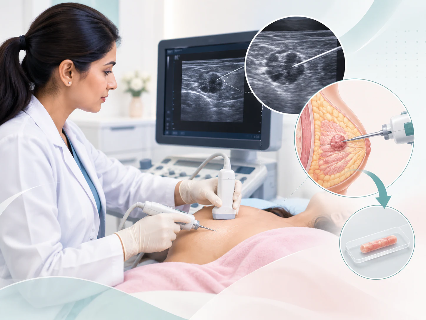
A core needle biopsy uses a needle to obtain small pieces of tissue from the area being assessed. These samples are sent for histopathological examination, where the tissue and its structure can be studied. The procedure samples the finding; it is not the same as an operation to remove the whole area.
Image guidance may help locate the intended target. The approach depends on how the finding is best seen and on the information needed. A technique name on a referral should be discussed in the context of the actual images, rather than treated as a choice patients must make themselves.
The illustration explains the concept, but it is not a set of procedural instructions. Positioning, preparation and sampling are planned by the clinical team for the individual target.
The clinical setting
Choosing guidance for the area being assessed
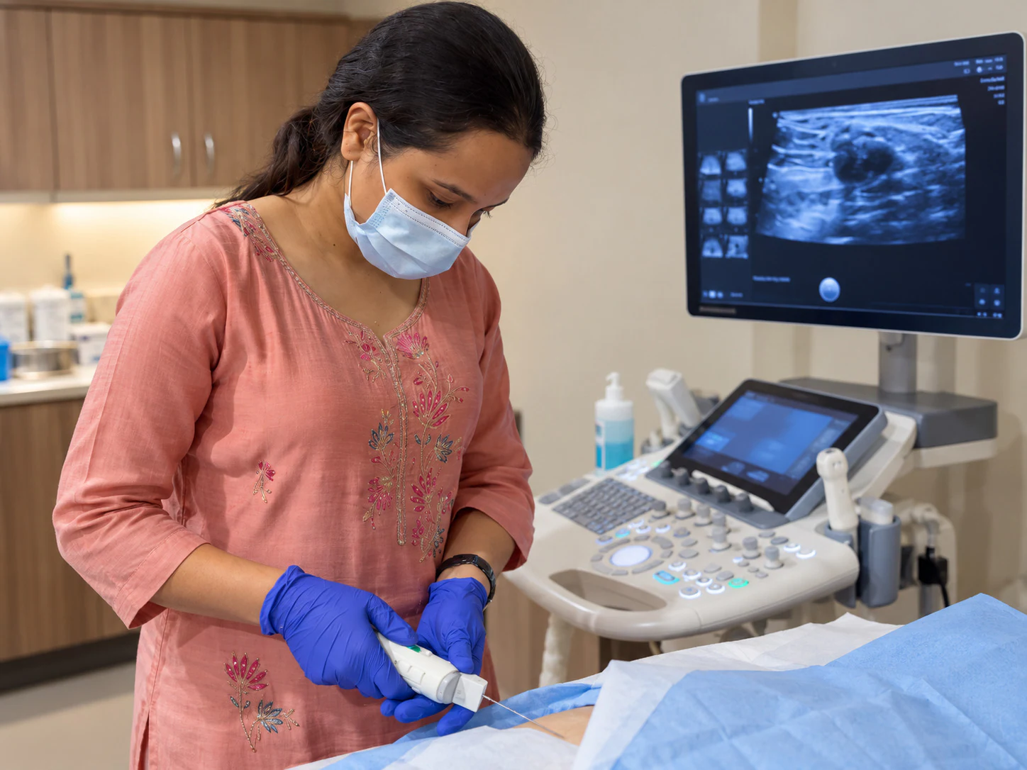
When a finding is visible on ultrasound and this approach is appropriate, real-time imaging can guide sampling. The reason for a biopsy and the method used to guide it are related but separate decisions. The team first needs to understand the target and then choose a suitable way to assess it.
Bring the earlier imaging that led to the recommendation. The radiologist may review the area again before proceeding. This review helps connect the referral, the current examination and the intended sample. If the planned approach changes, ask what has changed and why.
The photograph shows Dr Pranjali Joshi in a real image-guided procedure setting. It does not identify a particular sampling technique or imply that every patient has the same procedure.
Before the procedure
Bring your records and discuss your medicines
Bring the referral if available, previous breast images and written reports, and records of any earlier breast surgery or sampling. Include relevant pathology results. The images themselves may provide useful detail beyond the wording of a report, particularly if the assessment took place elsewhere.
Tell the team about relevant medical conditions, medicines, supplements and allergies, including any previous reaction to local anaesthetic. Mention a possibility of pregnancy where relevant to the planned assessment. Do not stop or change prescribed medicines independently: ask for instructions that apply to your own procedure.
Before travelling, confirm the requested procedure and any preparation requirements. Write down the questions you would like answered, including what the sample is intended to clarify and how results will be discussed. This page does not set a procedure duration or promise a reporting time.
For a breast biopsy enquiry in Lucknow, share the referral details and ask which earlier images and reports to bring. The team must confirm the sampling method, guidance and preparation for your finding.
A guided sequence
What happens during the visit?
The visit includes review of the imaging and confirmation of the area requiring assessment. The procedure should be explained, with an opportunity to discuss its purpose, relevant risks and alternatives before consent. Tell the team if you are anxious or have difficulty with the position needed.
Local preparation and pain relief are discussed as part of the procedure; local anaesthetic is commonly used for core sampling. You may still notice pressure or discomfort, so communicate how you are feeling. There is no promise that a procedure will be painless, and comfort needs differ between patients.
Review and positioning
The team reviews the target and helps you into a position suitable for the planned sampling.
Preparation and sampling
The area is prepared and samples are obtained using the guidance appropriate to the finding.
Sample handling and instructions
The material is prepared for laboratory examination. Before leaving, you receive instructions for care of the procedure site and the next steps.
After the visit
Follow the instructions given for your procedure
Mild local tenderness or bruising may occur after sampling, but what to expect depends on the procedure and the individual. Follow the specific instructions you receive about the dressing, activity and any medicines. General website information cannot replace advice based on how your procedure went.
Before leaving, make sure you know how to contact the treating team if you are concerned. Seek advice about unexpected or worsening symptoms, such as increasing swelling, ongoing bleeding or signs of infection. Do not assume a symptom is harmless simply because some local effects can occur after sampling.
Keep the aftercare information with your records. If any instruction is unclear, ask for it to be explained rather than trying to infer an activity restriction or medication plan from another patient’s experience.

Connecting the information
From an imaging finding to a next-step discussion
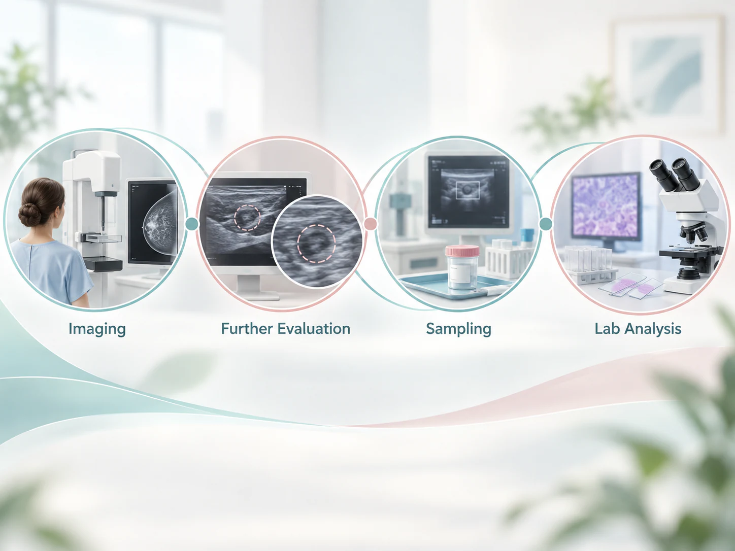
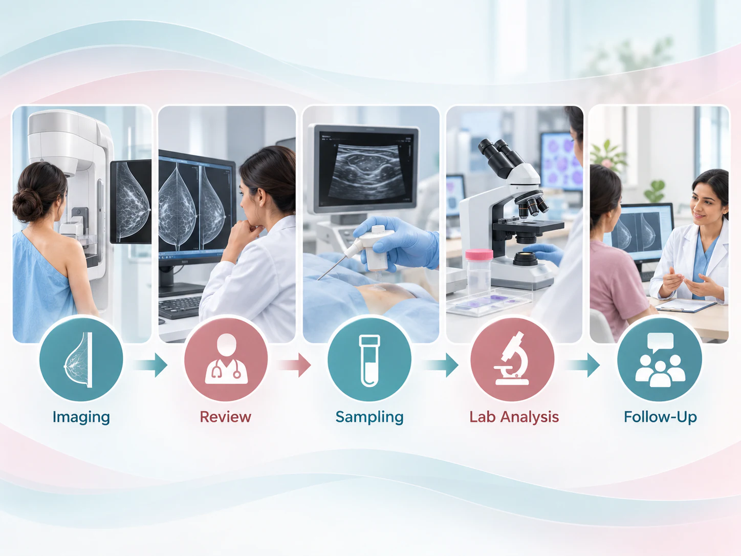
The biopsy is one stage in a wider assessment. The laboratory examines the tissue and provides a pathology report. That report is then considered alongside the imaging question and the clinical history. The important issue is whether the sample result helps explain the area that prompted the biopsy.
This is often described as imaging–pathology correlation. A report can contain unfamiliar language, and its meaning should be discussed in relation to your own assessment. Do not try to reach a conclusion from a single term without the accompanying recommendation.
Finding and sample
The area requiring clarification is identified and tissue is obtained for examination.
Laboratory and review
Pathology information is reviewed with imaging and clinical findings before the next step is discussed.
Results in context
What might happen after the report?
The next step depends on the result and how well it fits the imaging and clinical picture. Follow-up, additional imaging, further sampling or a treatment discussion may be recommended where appropriate. A sample does not guarantee that every question will be answered on the first occasion.
Ask how you will receive the written report and which member of your care team will explain it. If results from different examinations appear not to agree, that needs clinical review rather than choosing whichever wording sounds most reassuring. Keep copies so the information is available at subsequent appointments.
If an additional step is advised, clarify its purpose and who will coordinate it. No reporting deadline is promised here; obtaining the report and understanding the plan both matter.
Different kinds of sample
Core biopsy and FNAC answer different questions
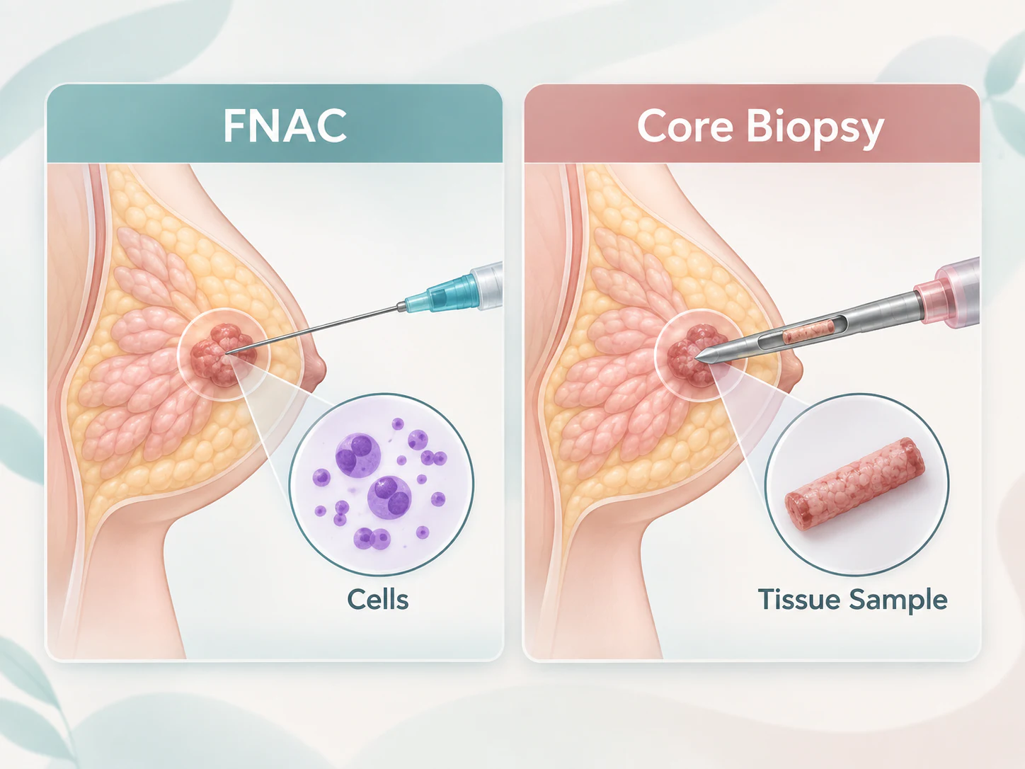
FNAC generally obtains cells or fluid for cytological assessment. Core biopsy obtains small tissue samples for histopathological assessment. The kind of information required, the imaging appearance and the clinical context help determine which method is appropriate.
Neither method should be selected from a website comparison alone. A recommendation for one approach does not mean the other is always inferior. Ask why the proposed sample is suited to the question your team is trying to answer.
Your questions
Common questions about breast biopsy in lucknow
Why might a breast biopsy be recommended?
It may be recommended when imaging or clinical assessment needs tissue information to clarify a finding. Ask which area is being sampled and what question the result is expected to answer.
Does a biopsy recommendation mean cancer?
No. The recommendation is a request for more information, not a diagnosis. The tissue result is considered with the imaging and clinical findings before next steps are discussed.
What is a core needle biopsy?
It is a procedure that obtains small tissue samples using a needle. The samples are examined in a laboratory, providing tissue information that can be considered alongside the imaging assessment.
Is breast biopsy the same as FNAC?
Core biopsy and FNAC obtain different types of material. Core biopsy provides tissue for histopathology; FNAC generally provides cells or fluid for cytology. The clinical team selects the method for the information needed.
How is the biopsy area located?
The approach depends on how the target is best seen. Ultrasound can guide selected procedures when the finding is visible and that form of guidance is appropriate. The team reviews the imaging before proceeding.
Do I need to bring previous breast images?
Bring actual images and reports if available, along with your referral and records of earlier procedures. These help connect the current recommendation with previous assessments.
Should I stop medicines before biopsy?
Do not stop prescribed medicines independently. Tell the team what you take, including supplements, and discuss allergies and relevant medical history. Follow the individual instructions provided for your procedure.
What happens after the procedure?
You receive aftercare instructions and information about the results process. Follow the advice for your own procedure and contact the treating team if you are concerned about symptoms afterwards.
What happens to the tissue sample?
It is sent for pathological examination. The laboratory report supplies information about the sample; it is not an instant result and should be reviewed with the reason for the biopsy.
How are biopsy results considered with imaging?
The team considers whether the pathology findings explain the area seen on imaging and fit the clinical picture. Further review, follow-up or additional sampling may be needed if a question remains unresolved.
Your next step
Prepare for your breast procedure
Bring your referral, earlier images and questions to the assessment. Dr Pranjali Joshi’s breast imaging and interventions practice in Lucknow considers sampling in the context of the finding being investigated. Understanding the purpose, aftercare and results plan can help you prepare for the next step.